What is Geographic Atrophy (GA)?
Understanding the eye
Your eyes help you navigate the world and connect with the people and things you love.
Vision happens when your eyes turn light into signals
your brain can recognize.
Learning about how the eye works will help you understand how eye diseases like Geographic Atrophy (GA), an advanced form of dry age-related macular degeneration (AMD), affect the eye.
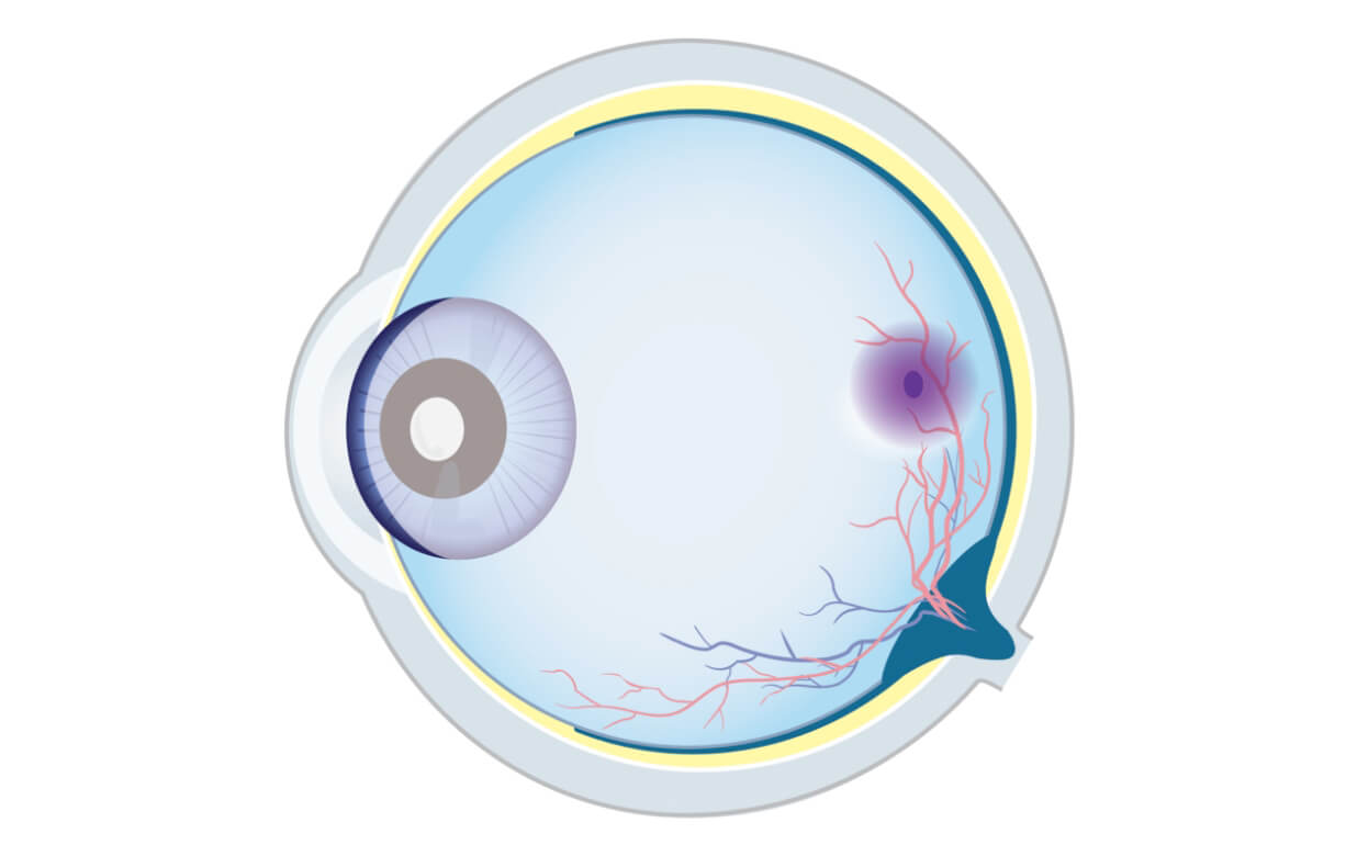
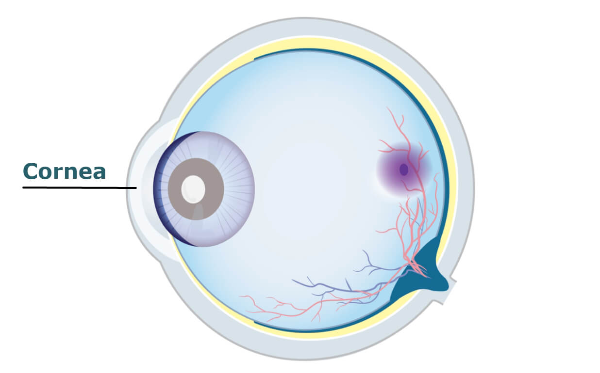
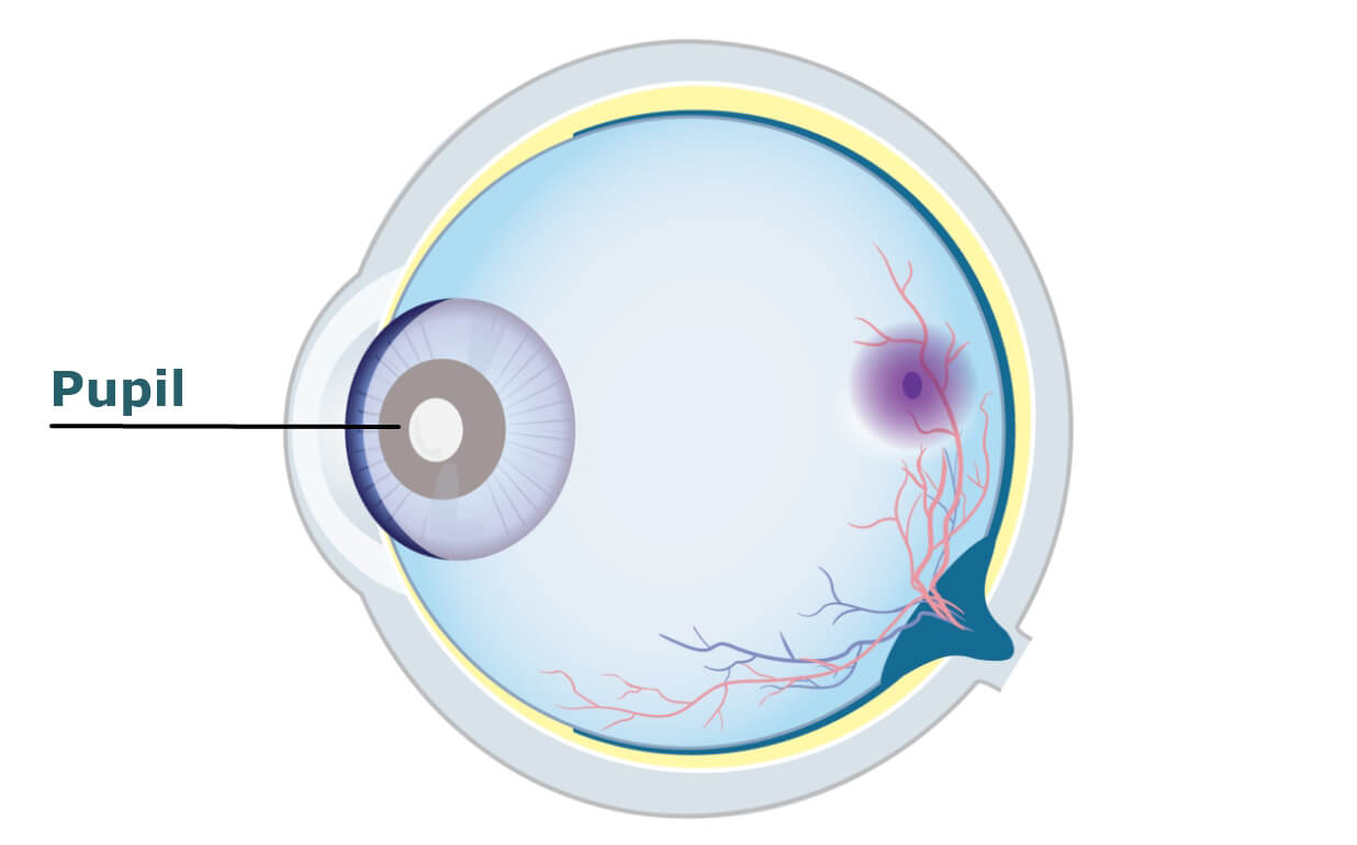


Images are for illustrative purposes only. Drawing is not a true cross-section.
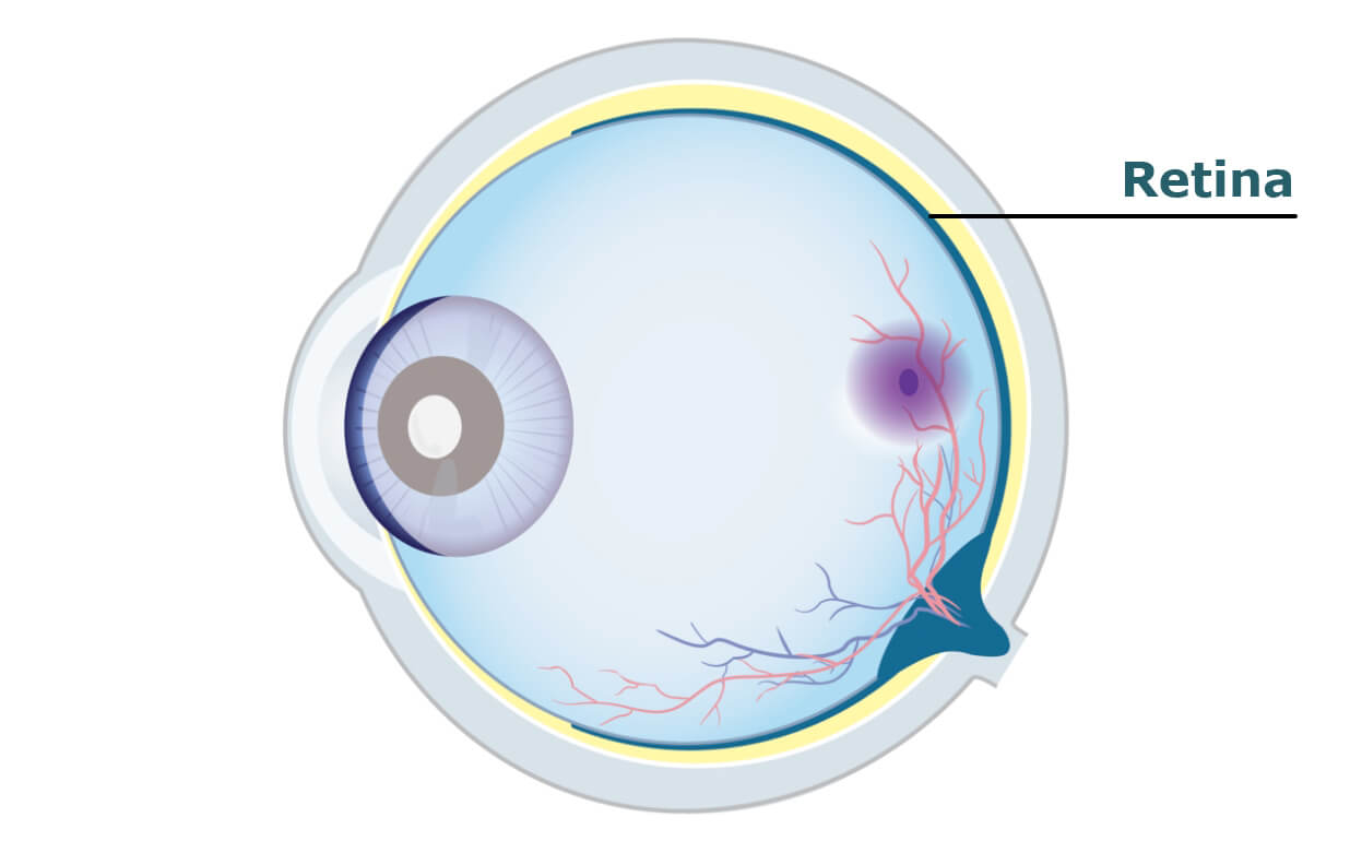
GA is a progressive disease that affects the retina, specifically the macula. Damage to these areas of the eye can lead to permanent vision loss.
AMD and GA
AMD is an eye disease primarily found in people over the age of 50. When you have this condition, parts of your macula begin to thin.
Early/intermediate AMD can lead to GA.
Recognizing the differences between early, intermediate, and advanced AMD (wet AMD and GA) can help you have better conversations with your eye doctor. It’s important to know that each of your eyes can be different. One eye may have AMD symptoms while the other may not.
Click on the tabs below to learn more about each eye condition.
- With early or intermediate AMD, you may not have symptoms at all, or symptoms like blurriness and difficulty seeing in low light may be mild
- As early or intermediate AMD gets worse, it causes reduced vision and blurriness in the areas you are trying to focus on. Straight lines may also appear crooked
- When you have AMD, even small changes to your vision can mean your disease is getting worse
Early/intermediate AMD can lead to GA, wet AMD, or both GA and wet AMD
Images are for illustrative purposes only. Drawing is not a true cross-section.
GA can develop as AMD progresses and becomes more advanced.
- It affects the retina – a thin layer of tissue lining the back of the eye that senses light, allowing you to see
- In GA, cells in the retina start to die, or atrophy; when your eye doctor looks at your retina, these regions of dead and dying cells look like areas on a map, which is why it’s called “Geographic Atrophy”
- GA can lead to permanent vision loss, and a blurry spot in the centre of your vision that can make activities like reading and driving difficult, especially in low light or dark places
- GA is progressive and irreversible – vision changes due to GA get worse over time and cannot be corrected with glasses or through surgery
Images are for illustrative purposes only. Drawing is not a true cross-section.
- Wet AMD shares similar vision changes to those seen with GA, including blurred vision and reduced central vision
- It gets a “wet” distinction as abnormal blood vessels may cause fluid or blood to leak into the macula
Images are for illustrative purposes only. Drawing is not a true cross-section.
Images are for illustrative purposes only. Drawing is not a true cross-section.
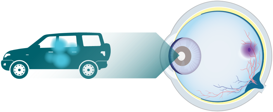
How GA impacts vision
GA is a disease that gets worse over time and can cause permanent vision loss. Talking to your eye doctor about changes in vision can help in getting an early diagnosis.
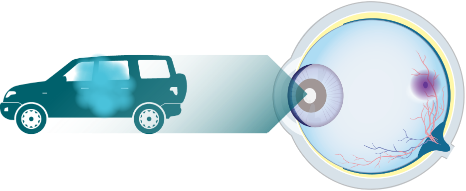
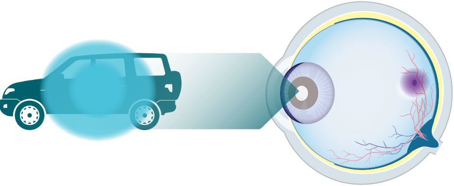
Hover over the images to see some signs and symptoms of GA:
Difficulty seeing in the dark


Hazy or blurred vision (less sharp or detailed)


Straight lines appear crooked

A small but growing blurry spot in the centre of vision


Colours seem dull or washed out
Images are for illustrative purposes only. Vision impairment due to GA may vary.
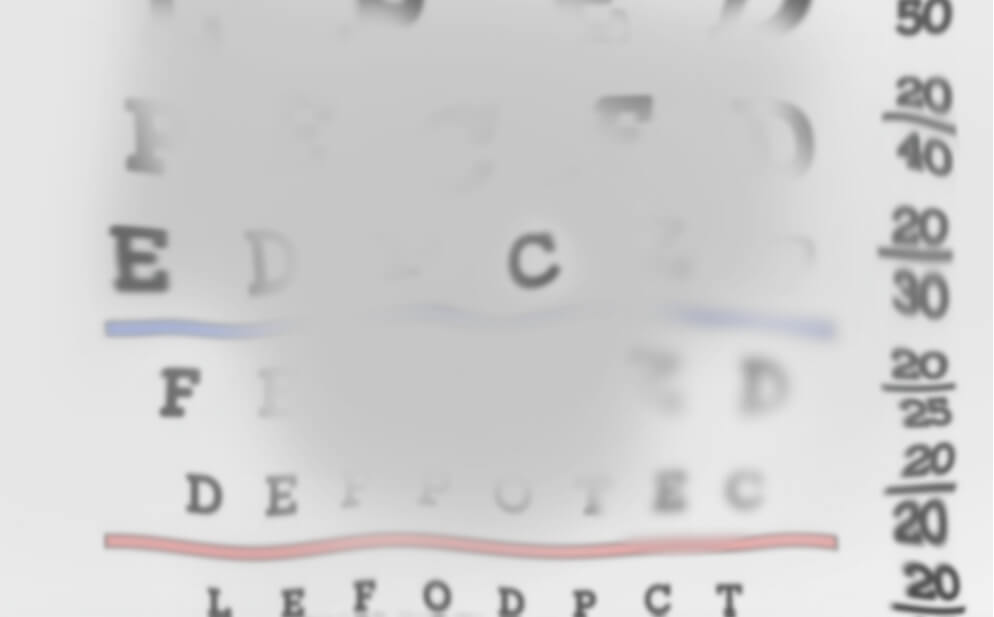
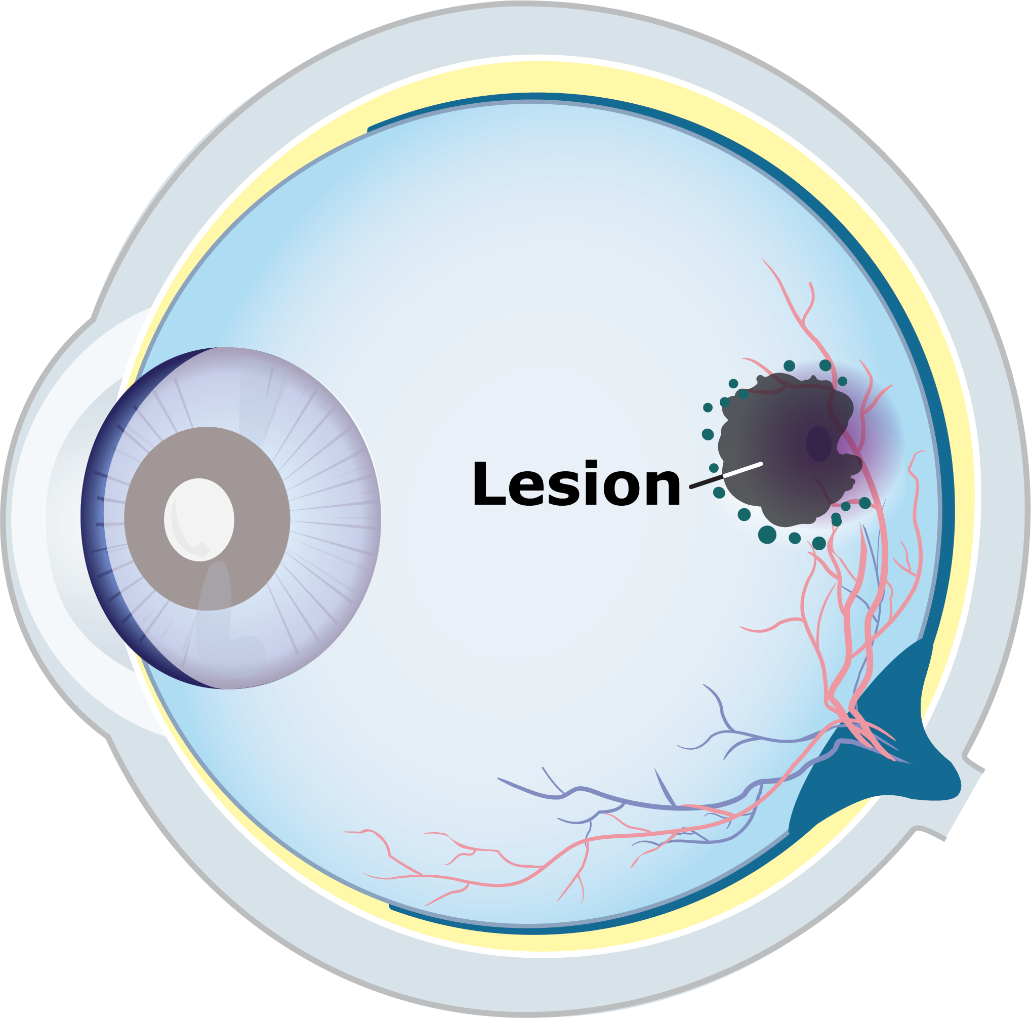
With GA, you may not notice changes in your vision when you look at an eye chart. However, reading and driving at night may become harder as patches of damage (also known as lesions) grow larger.
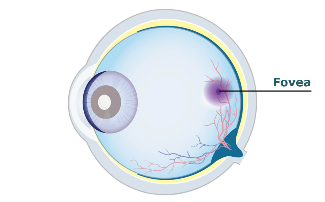
The fovea is the part of the eye that allows you to read and drive.
In a study of 397 patients with GA who had lesions impacting the central part of the retina (the fovea), those lesions reached the fovea in a median of just 2.5 years from diagnosis.*
AMD=age-related macular degeneration; GA=Geographic Atrophy.
*Part of a long-term study of 3640 patients with signs of early or more advanced forms of AMD.
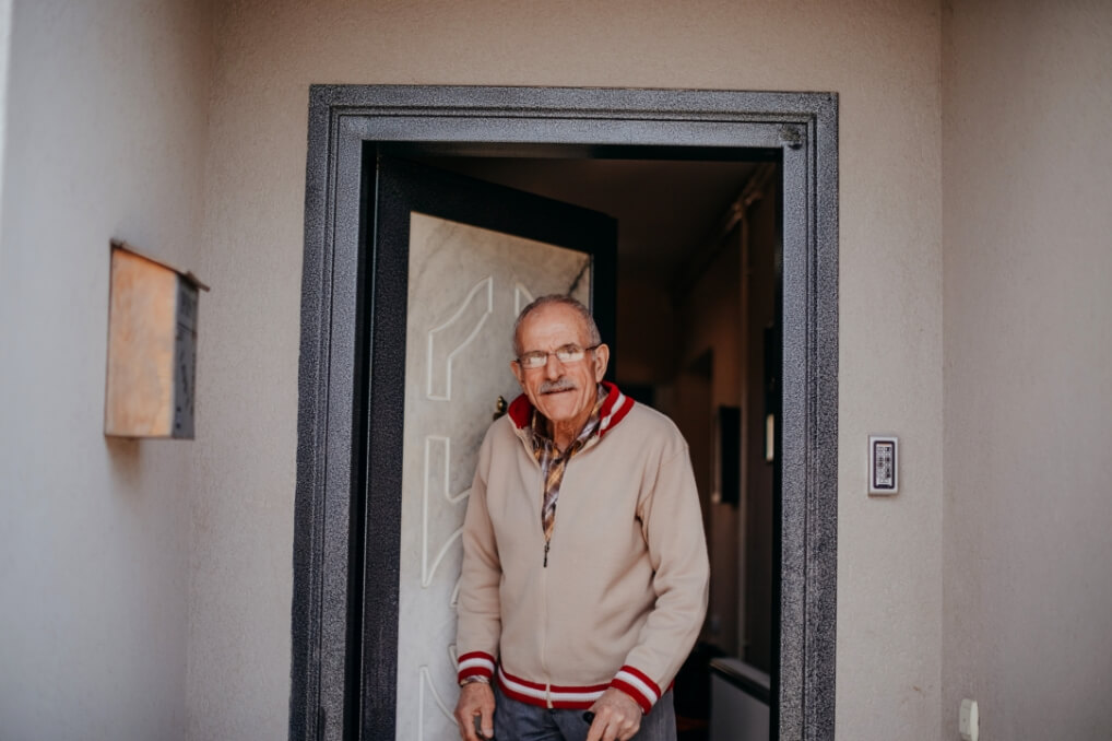
See how GA changes vision over time
Click to experience the progression of GA over time.

For illustrative purposes only. Vision impairment due to GA may vary.
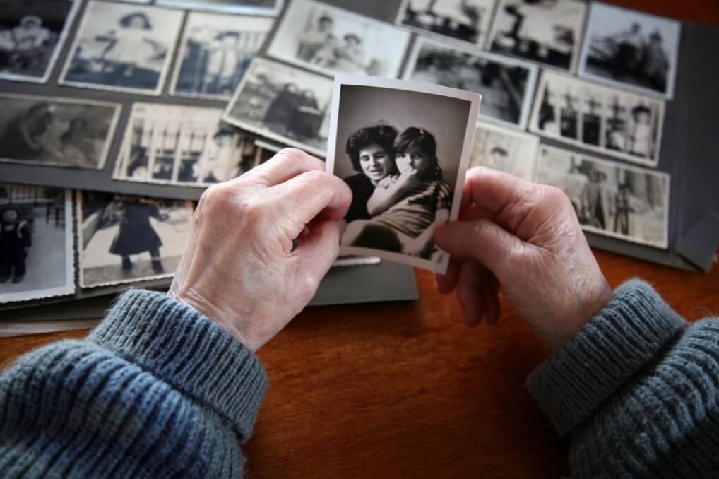
At diagnosis
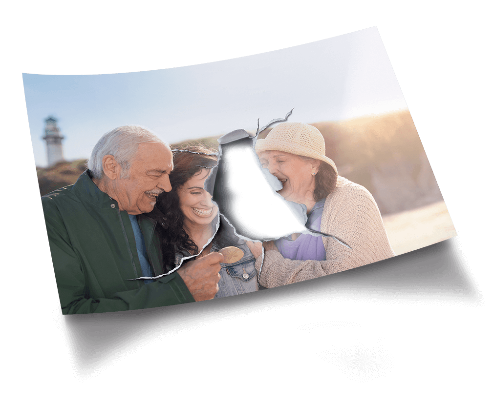
This image shows the vision of a 70-year-old woman who has just been diagnosed with GA in both eyes. Her eyes have multiple lesions in the area surrounding the fovea, making it difficult for her to see the faces of her loved ones.

For illustrative purposes only. Vision impairment due to GA may vary.
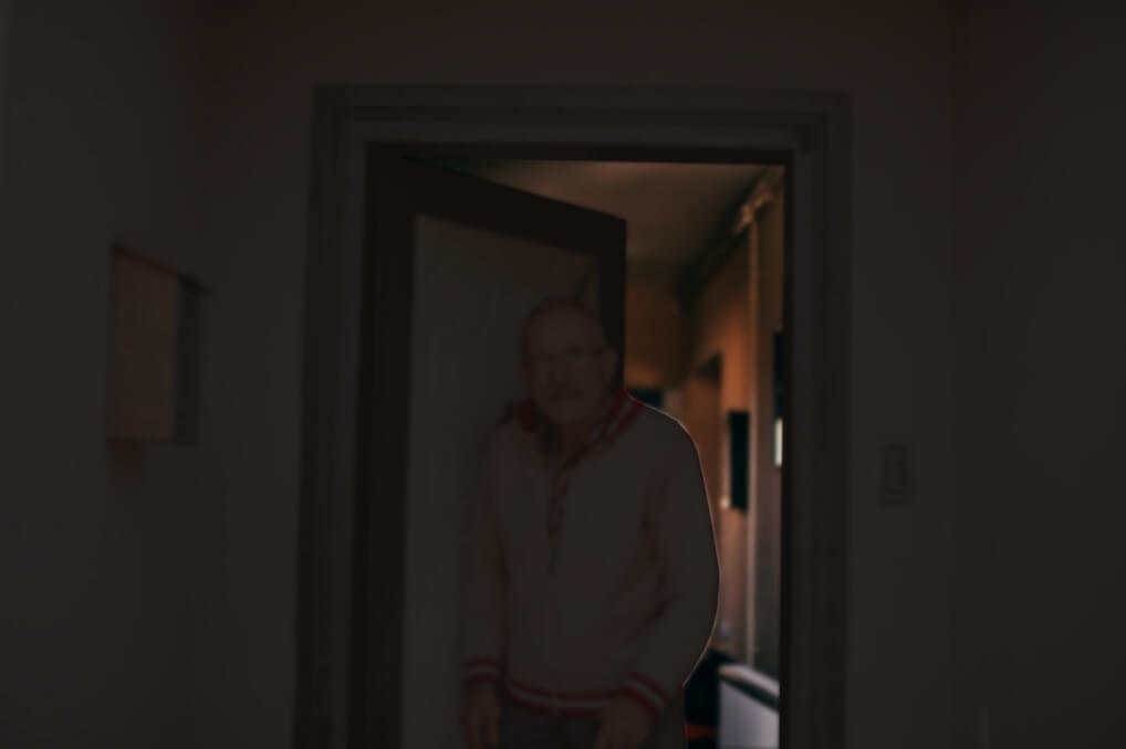
+2 years
GA has progressed in both of the patient’s eyes. The faces of loved ones have become harder to see. Driving and other daily activities may become unsafe as lesions grow larger.
For illustrative purposes only. Vision impairment due to GA may vary.
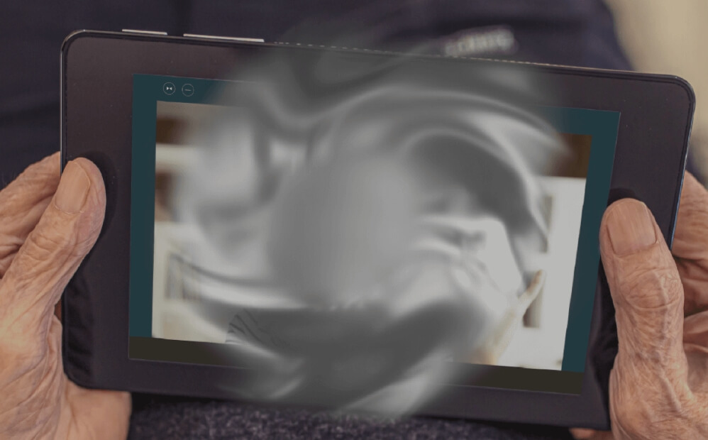
+6 years
Now, with increased progression in both eyes, the faces of loved ones, the ability to live independently, and overall quality of life have all been impacted by vision loss due to GA.

For illustrative purposes only. Vision impairment due to GA may vary.
At diagnosis
This image shows the vision of a 70-year-old woman who has just been diagnosed with GA in both eyes. Her eyes have multiple lesions in the area surrounding the fovea, making it difficult for her to see.

For illustrative purposes only. Vision impairment due to GA may vary.
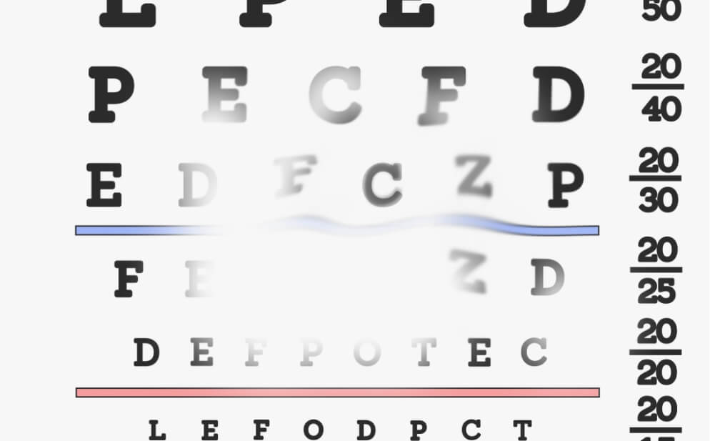
+2 years
GA has progressed in both of the patient’s eyes. Reading has become more difficult. Driving and other daily activities may become unsafe as lesions grow larger.

For illustrative purposes only. Vision impairment due to GA may vary.
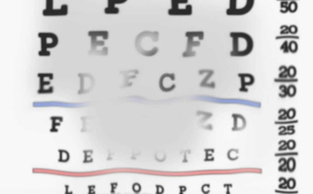
+6 years
Now, with increased progression in both eyes, reading, the ability to live independently, and overall quality of life have all been impacted by vision loss due to GA.
For illustrative purposes only. Vision impairment due to GA may vary.
At diagnosis
This image shows the vision of a 70-year-old woman who has just been diagnosed with GA in both eyes. Her eyes have multiple lesions in the area surrounding the fovea, making it difficult for her to see.
For illustrative purposes only. Vision impairment due to GA may vary.
+2 years
GA has progressed in both of the patient’s eyes. Letters have become harder to see. Driving and other daily activities may become unsafe as lesions grow larger.
For illustrative purposes only. Vision impairment due to GA may vary.
+6 years
Now, with increased progression in both eyes, reading, the ability to live independently, and overall quality of life have all been impacted by vision loss due to GA.
Be prepared to talk to your eye doctor about changes in vision and potential signs of GA.
GA in focus
Watch this video to learn more about what’s going on in your eye when you have GA.

Close your eyes.
Let them adjust.
Now, open them.
Notice how the world around you gets clearer as the light pours in?
Your eyes help you navigate the world and connect you with the people and things you love, so it’s important to understand what’s happening if you experience vision changes.
Geographic Atrophy, or GA, is an advanced form of age-related macular degeneration, or AMD.
This condition can lead to permanent and irreversible vision loss.
If you’re early in your GA journey, the effects may not be noticeable now. But over time, details could start to look hazy or blurred.
Many things can increase your risk of getting GA, such as your age and lifestyle.
Your family history can make up to seventy percent of your risk of getting GA.
To understand how GA happens, I want you to think about the palms of your hands.
When you’re able to see all the crisscrossing lines and details, it’s because of a part of your eye called the retina.
It turns light – and all of the colours and the shapes it captures – into signals that your brain can understand.
Your retina is located in an area at the back of your eye.
Within your retina is a tiny section called the macula. It helps you to process all the fine details of your world clearly.
It’s the retina – and more specifically the macula – where GA affects your vision the most.
When you have GA, excessive debris deposits called drusen can build up in your retina and lead to the damage of cells.
While this damage may start small, it will progress. As cells continue to die off, larger patches known as lesions can grow and spread across the retina.
If you’ve been living with GA, you might notice that driving at night and reading have become harder.
Straight lines, like telephone poles or those on a piece of paper, may have begun to look wavy or distorted. These are all signs that GA is progressing.
As GA continues to progress, you may notice a blurry spot that appears in the centre of your vision.
As the lesions caused by GA grow larger, its effect on vision worsens, and can eventually lead to permanent vision loss.
If you are experiencing symptoms of GA, know that you aren’t alone: GA affects approximately five million globally.
Finding resources and a supportive community will be an important step in your GA journey.
Play an active role and stay involved with your vision to help you adapt to life with GA.
Every person’s experience with GA is unique. Speak with your doctor about questions you may have regarding GA and its symptoms.
How GA develops in the eye
Risk factors
Family history (including genetics), aging, history of cigarette smoking, high blood pressure, obesity, low physical activity, and UV exposure can increase your risk of getting AMD, which may lead to GA.
Drusen
These risk factors can lead to a buildup of debris in the eye’s retina. This debris is called drusen and is made up of proteins and fats that the body no longer needs.
Overactivation of the immune system and inflammation
This buildup of drusen can contribute to an overactivation of parts of the immune system in the eye.
This leads to inflammation, which contributes to your body mistakenly damaging and destroying healthy cells in a part of your retina called the macula.
While this damage in the eye may start small, cells continue to die off and form larger patches, known as lesions, that get bigger and spread across the retina.
As these lesions grow larger, GA worsens and can cause permanent vision loss.
Risk factors
Drusen
Overactivation of the
immune system and
inflammation
Cell death
Lesion growth
Vision loss
How GA progresses in the eye
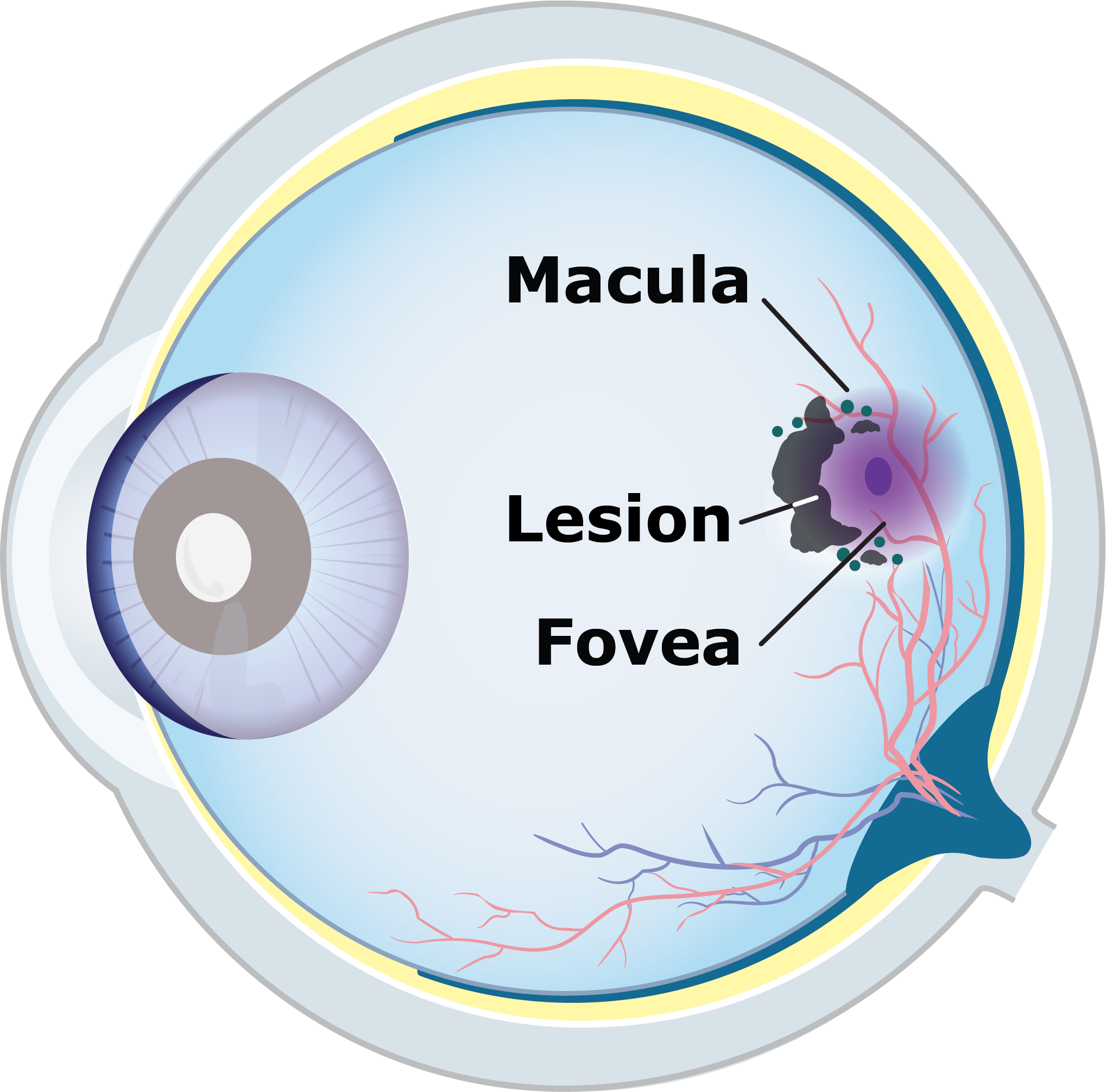
Start of GA
Damaged cells in the macula begin to die off, forming areas in the retina called lesions.
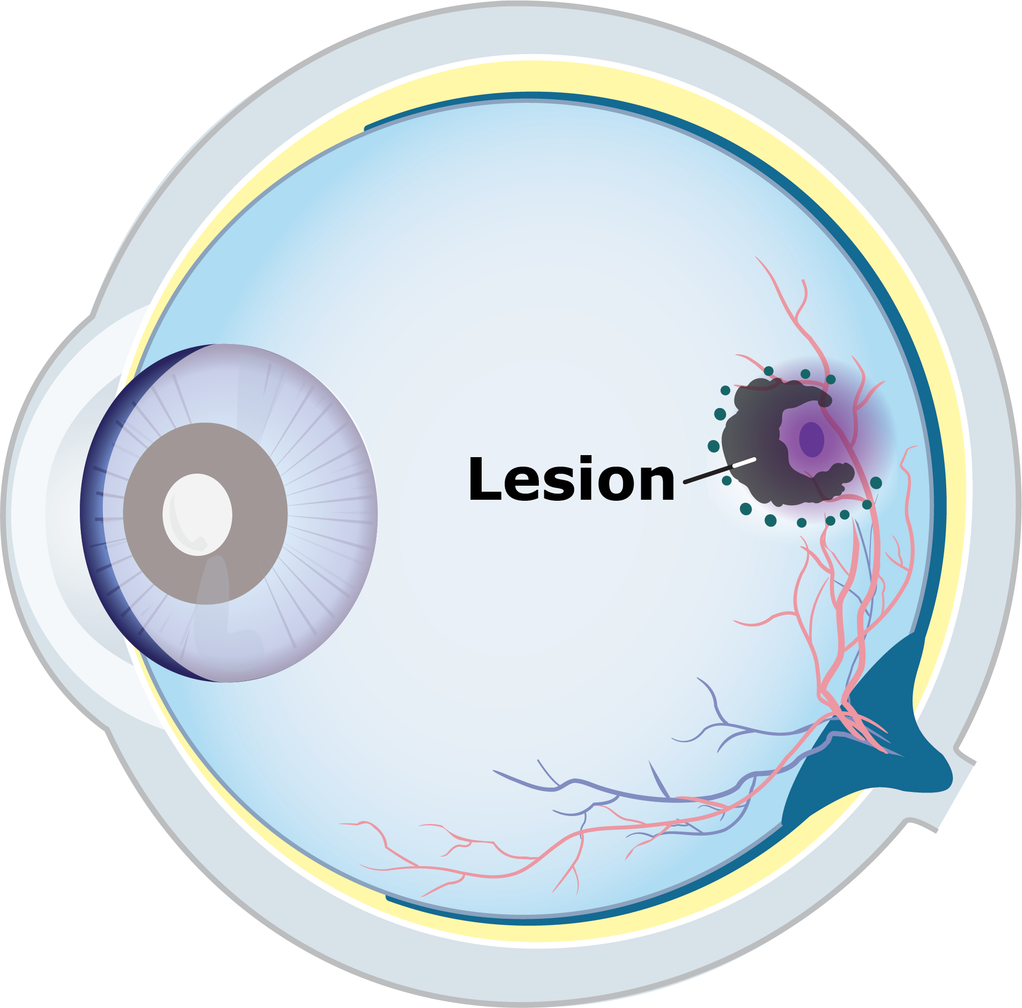
GA lesion growth
As damage continues to progress, lesions can grow larger and spread across the retina.
Foveal damage
Over time, lesions reach the fovea at the centre of the macula, causing loss of central, detailed vision.
Contributing factors for GA
Below is a list of things that may contribute to GA:
Age
Family history (the genes you inherit play ~70% of the role in GA development)
Diet high in fatty foods
High blood pressure
History of smoking
Low physical activity
Obesity
Learn more about GA